Fig. 1.
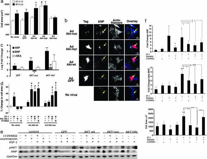
Akt-nuc inhibits hypertrophy through an ANP-dependent signaling cascade. (a) Akt-nuc inhibits endothelin-induced hypertrophy. Cell surface area measurements are from cardiomyocytes that were either uninfected (No virus) or infected with adenoviruses expressing GFP (Ad GFP), wild-type Akt (Ad Akt-wt), myristoylated Akt (Ad Akt-myr), or nuclear-targeted Akt (Ad Akt-nuc). Each cell population was measured with infection alone (white bars) or infection followed by 48-h stimulation with hypertrophic agonist ET-1 (10−7 mol/liter) (black bars). ∗, P < 0.05 vs. same virus without ET-1. #, P < 0.05 vs. no virus without ET-1. †, P < 0.05 vs. no virus with ET-1. (b) Confocal microscopy of cardiomyocytes stimulated with ET-1 (10−7 mol/liter) indicates that Akt-nuc promotes ANP expression. Antibodies to myc-tag, hemagglutinin-tag, or GFP-tag show infected cells (Tag), whereas ANP expression was detected by anti-ANP antibody (ANP). Tag (red), ANP (green), and actin (blue) are depicted in overlay images. (c) Akt-nuc induces ANP without concomitant increases in α-skeletal actin (αSKA). ANP mRNA transcript levels are significantly increased relative to GFP-expressing control (black bars; ∗∗, P < 0.01). BNP expression shows differential and significant changes between Akt-nuc- and Akt-myr-expressing cells (gray bars; #, P < 0.05; ##, P < 0.01). Expression of hypertrophic marker αSKA is not increased by Akt-nuc expression but is significantly elevated by Akt-myr expression (white bars, ++, P < 0.01). (d) Antihypertrophic effects of Akt-nuc involve autocrine and/or paracrine stimulation of GC-A receptor and PKG. Hypertrophy of cardiomyocytes induced by ET-1 (10−7 mol/liter) was inhibited in Ad Akt-nuc infected cells. Antihypertrophic effects of Ad Akt-nuc were reversed by inhibition of GC-A receptor with HS-142-1 (10 μg/ml) or PKG with KT5823 (10−6 mol/liter). ∗, P < 0.05 vs. no virus without any drugs. #, P < 0.05 vs. Ad Akt-wt without any drugs. †, P < 0.05 vs. Ad Akt-nuc without any drugs. §, P < 0.05 vs. Ad Akt-nuc with ET-1. (e) IGF-mediated induction of ANP depends upon PI3-K/Akt signaling. Immunoblot analysis of cultured cardiomyocyte lysates infected with adenoviral vectors expressing GFP, Akt-wt, Akt-nuc, or Akt-DN proteins. Cultures were subsequently treated the next day with IGF-1 alone or in combination with PI3-K inhibitors LY294002 or wortmannin. (f) Antihypertrophic effect of estradiol is abrogated by inhibition of PI3-K. Cultured cardiomyocytes were exposed to estradiol alone or in combination with LY294002 and subsequently stimulated by ET-1 treatment. ∗, P < 0.05; ∗∗, P < 0.01 for each comparison indicated. See Supporting Text, which is published as supporting information on the PNAS web site, for expanded detailed information of this figure.

