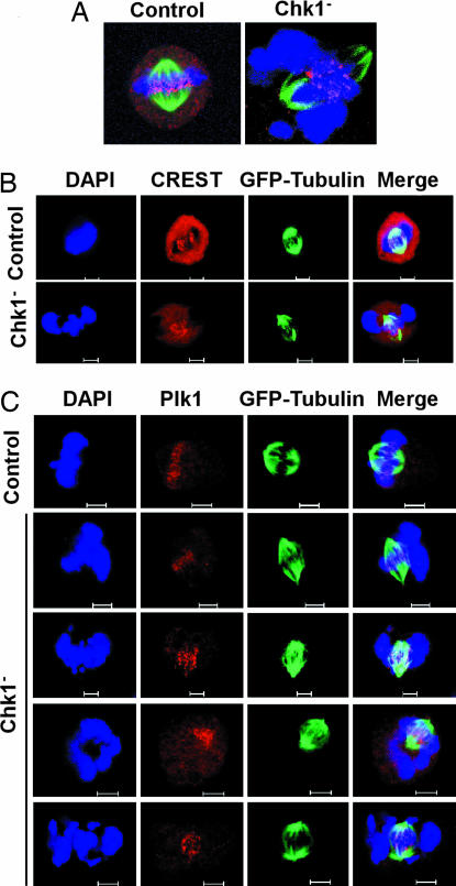
Fig. 4.
Kinetochore defects in Chk1-depleted cells. HeLa cells stably expressing GFP-tubulin were Chk1-depleted by using the protocol described in Fig. 2C, released for 14 h, and stained with antibodies as indicated. DNA was stained with DAPI. (A) Representative images of Hec1 (red) antibody staining of control and Chk1-depleted HeLa cells. A series of Z-stack images was taken from mitotic cells; projections of the total Z-stack are shown. (B and C) Representative images of anti-CREST (B) or anti-Plk1 (C) staining patterns of control and Chk1-depleted cells during metaphase. (Scale bar, 5 μm.)

