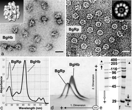
Fig. 1.
Identification of purified BgHb and BgRp. (a) Electron microscopy of HPLC-purified BgHb molecules. (a Inset) Three-dimensional reconstruction (see Fig. 5). (b) Electron microscopy of HPLC-enriched BgRp molecules. (b Inset) Class sum image from 10 top views aligned by IMAGIC. (Scale bars: 25 nm.) (c) UV-visible spectra of BgHb and BgRp. (d) Tandem-crossed immunoelectrophoresis of purified BgHb and BgRp against rabbit antibodies versus B. glabrata hemolymph proteins. Note the separate precipitation of the two protein peaks indicating nonidentity. (e) SDS/PAGE of BgHb (arrow) showing an apparent Mr of 180 kDa. A trace of the disulfide-bridged dimer of BgHb is also visible (arrowhead). Marker protein masses are indicated in kilodaltons. (f) SDS/PAGE of BgRP, showing two polypeptides (arrows) with 31 and 25 kDa, respectively. Traces of the hemocyanin subunit (arrowhead; see also Fig. 2) are also visible.

