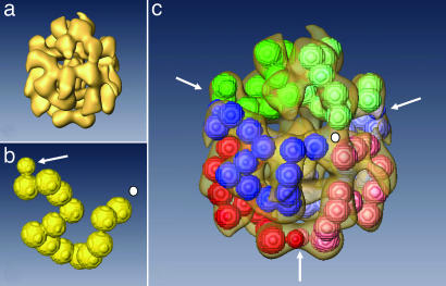
Fig. 5.
Three-dimensional reconstruction of the quaternary structure of BgHb. (a) 3D reconstruction of native BgHb (resolution, 3 nm). (b) Model of the BgHb polypeptide subunit with 13 globular masses for the heme domains and N-terminally a smaller mass for the plug domain (arrow). (c) The same view as in a, incorporating six polypeptide subunits as modeled in b, distinguished by different colors. By bringing together the N-terminal plug domains (arrows) of pairs of polypeptide chain subunits, this model is consistent with the occurrence of disulfide-bridged dimers of polypeptide chains, as was demonstrated biochemically. Additionally, subunits form triplets by joining C-terminal domains (white dot). Also see Fig. 6.

