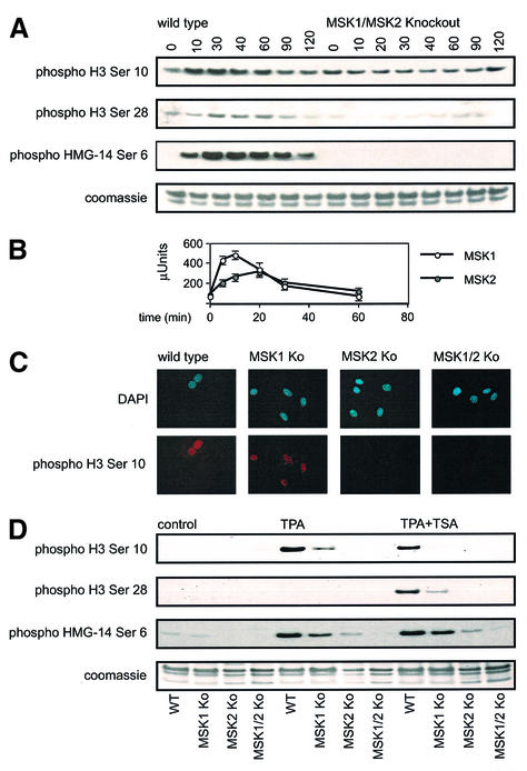
Fig. 6. Phosphorylation of histone H3 and HMG-14 in MSK1/MSK2 knockout cells by TPA. (A) Wild-type and MSK1/MSK2 knockout fibroblasts were serum starved overnight and then stimulated with TPA (400 ng/ml) for the times indicated. Cells were lysed and analysed for TPA-induced phosphorylation of histone H3 on Ser10 and Ser28 and of HMG-14 on Ser6 by immunoblotting. (B) MSK1 and MSK2 were assayed against Crosstide after immunoprecipitation from 0.5 mg of soluble protein lysate from wild-type cells. One unit of activity was defined as the amount of enzyme required to incorporate 1 nmol of phosphate into the substrate in 1 min. Six measurements were taken per time point, and error bars represent the SEM of these values. (C) Wild-type, MSK1, MSK2 and MSK1/MSK2 knockout fibroblasts were serum starved overnight and then stimulated with 400 ng/ml TPA for 30 min. Cells were fixed and phosphorylation of histone H3 on Ser10 examined by immunofluorescence. DAPI staining of the nucleus is shown in blue, and phospho-Ser10 histone H3 staining is shown in red. (D) Wild-type, MSK1, MSK2 and MSK1/MSK2 knockout fibroblasts were serum starved overnight, followed by treatment with 500 ng/ml TSA for 4 h where indicated. Cells were then stimulated with TPA (400 ng/ml) for 30 min. Cells were lysed and analysed for anisomycin-induced phosphorylation of histone H3 on Ser10 and Ser28 and of HMG-14 on Ser6 by immunoblotting.

An official website of the United States government
Here's how you know
Official websites use .gov
A
.gov website belongs to an official
government organization in the United States.
Secure .gov websites use HTTPS
A lock (
) or https:// means you've safely
connected to the .gov website. Share sensitive
information only on official, secure websites.
