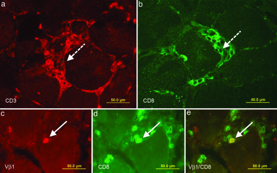
Fig. 2.
Immunolocalization of T cells in muscle tissue of patient PM16488. Cryosections of a frozen biopsy specimen were stained in red with a Cy3-labeled antibody to CD3 (a) and in green with an FITC-labeled antibody to CD8 (b). Dense focal invasions of T cells into muscle fibers are indicated in both figures by dashed arrows. Focal invasions are one of the diagnostic criteria of PM. However, such closely packed T cell aggregates are not suited for the isolation of single T cells by microdissection. We therefore focused on distinct single cells that indent or directly contact a muscle fiber. (c–e) Double stainings of TCR Vβ1 and CD8. Biopsy sections were stained in red with a Cy3-labeled anti-Vβ1 antibody (c) and in green with an FITC-labeled anti-CD8 antibody (d). (e) A double-positive cell in direct contact with a muscle fiber appears in yellow and is indicated by an arrow.

