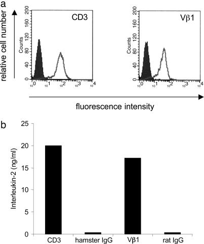
Fig. 3.
Characterization of the Vα2.2-Vβ1-TCR reconstituted in the T hybridoma cell line 58α−β−. (a) Cell-surface expression as determined by flow cytometry. The TCR transfectants were stained positive with antibodies to mouse CD3 (a Left) and human TCR Vβ1 (a Right) molecules (white areas). The transfectants were not stained with the corresponding isotype control antibodies hamster IgG and rat IgG, respectively (shaded areas). Clone A5 is shown as a representative of three independent clones. These data provide evidence that the reconstituted TCR is expressed at the cell surface of 58α−β− cells. (b) Antibody-induced activation of the reconstituted Vα2.2-Vβ1-TCR clone A5. Antibodies to mouse CD3, Vβ1, and the corresponding isotype control antibodies were coated on microtiter plates, the TCR transfectants were added, and IL-2 was measured in the supernatants by ELISA. The transfectants produced IL-2 in response to anti-CD3 and anti-Vβ1 antibodies but not to the respective control antibodies. These results show that the reconstructed, revived TCR is functional.

