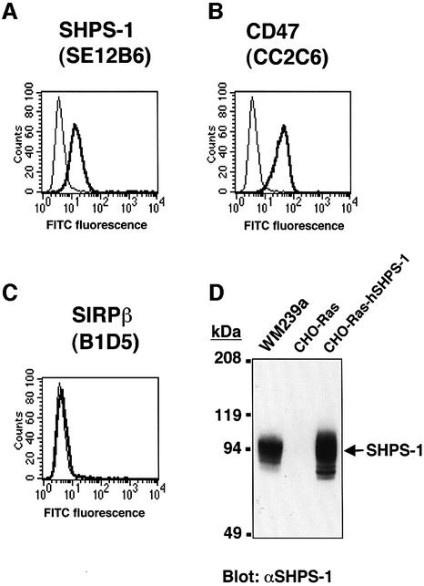
Fig. 1. Surface expression of SHPS-1 and CD47 on WM239a melanoma cells. Cells were incubated with the mAbs SE12B6 to SHPS-1 (A), the mAb CC2C6 to CD47 (B), or the mAb B1D5 to SIRPβ (C). Immunocomplexes were then detected with FITC-conjugated goat pAbs to mouse IgG (thick trace) and flow cytometry. The specific mAbs were replaced by normal mouse IgG as a negative control (thin trace). (D) Whole-cell lysates (20 µg of protein) of WM239a cells, of CHO-Ras cells (a negative control), or of CHO-Ras cells stably expressing human SHPS-1 (CHO-Ras-hSHPS-1 cells; a positive control), were subjected to immunoblot analysis with pAbs to SHPS-1 (αSHPS-1). The positions of SHPS-1 and of molecular size standards are indicated. All data are representative of three independent experiments.

An official website of the United States government
Here's how you know
Official websites use .gov
A
.gov website belongs to an official
government organization in the United States.
Secure .gov websites use HTTPS
A lock (
) or https:// means you've safely
connected to the .gov website. Share sensitive
information only on official, secure websites.
