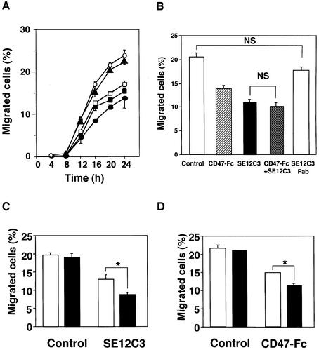
Fig. 2. Effects of SHPS-1 ligands on WM239a cell migration. (A) Cells were preincubated with mAbs SE12C3 (closed circles) or SE12B6 (open squares) to SHPS-1 (5 µg/ml), human CD47-Fc (closed squares; 10 µg/ml), mAb B1D5 (closed triangles) to SIRPβ (5 µg/ml) or control mouse IgG (open circles; 10 µg/ml) for 30 min at 37°C and were then applied to polycarbonate filters in the upper compartments of a Transwell apparatus. After incubation for the indicated times, the number of cells that had migrated into the lower compartments was determined and expressed as a percentage of the total cells applied. (B) Cells were similarly preincubated with control mouse IgG (10 µg/ml), mAb SE12C3 (5 µg/ml), human CD47-Fc (10 µg/ml), SE12C3 (5 µg/ml) plus CD47-Fc (10 µg/ml) or Fab fragments of mAb SE12C3 (5 µg/ml) and then subjected to assay of cell migration for 20 h. (C) Migration assays were performed for 20 h with WM239a cells with control mouse IgG (5 µg/ml) or with mAb SE12C3 (5 µg/ml), each either in the absence (open columns) or presence (solid columns) of goat pAbs to mouse IgG (10 µg/ml). (D) Migration assays were performed for 20 h with cells with control human IgG (10 µg/ml) or with human CD47-Fc (10 µg/ml), each either in the absence (open columns) or presence (solid columns) of goat pAbs to human IgG (10 µg/ml). Data in (A) are means ± SE of triplicates from an experiment that was repeated a total of three times with similar results. Data in (B), (C) and (D) are means ± SE of values from three independent experiments. NS, not significant, *P < 0.05 for the indicated comparisons (Student’s t-test).

An official website of the United States government
Here's how you know
Official websites use .gov
A
.gov website belongs to an official
government organization in the United States.
Secure .gov websites use HTTPS
A lock (
) or https:// means you've safely
connected to the .gov website. Share sensitive
information only on official, secure websites.
