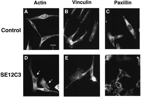
Fig. 6. Effects of SHPS-1 ligation on cytoskeletal architecture in WM239a cells. Cells cultured on chamber slides were incubated for 30 min at 37°C with control mouse IgG (5 µg/ml) (A–C) or the SE12C3 mAb to human SHPS-1 (5 µg/ml) (D–F). They were then washed, fixed and stained with rhodamine-conjugated phalloidin (A, D) or with mAbs either to vinculin (B, E) or to paxillin (C, F). Immunocomplexes were detected with FITC- conjugated secondary antibodies. Cells were then examined by fluorescence microscopy. Arrows indicate cells that exhibit a marked change in morphology in response to the SE12C3 mAb. Scale bar: 20 µm. Data are representative of three independent experiments.

An official website of the United States government
Here's how you know
Official websites use .gov
A
.gov website belongs to an official
government organization in the United States.
Secure .gov websites use HTTPS
A lock (
) or https:// means you've safely
connected to the .gov website. Share sensitive
information only on official, secure websites.
