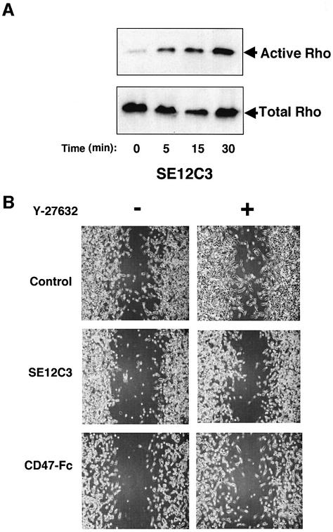
Fig. 7. Effects of SHPS-1 ligation on Rho GTPase activity (A) and reversal by Y-27632 of the SHPS-1 ligand-induced inhibition of cell migration (B). (A) Monolayers of WM239a cells at ∼90% confluence were incubated for the indicated times at 37°C with the SE12C3 mAb to human SHPS-l, after which the active form of Rho was precipitated from cell lysates with a GST fusion protein containing the Rho-binding domain of Rhotekin. The resulting precipitates were then subjected to immunoblot analysis with a mAb to RhoA (top panel). Whole-cell lysates were also directly subjected to immunoblot analysis with the same mAb to determine the total amount of Rho (bottom panel). (B) Monolayers of WM239a cells were preincubated with (right panels) or without (left panels) 10 µM Y-27632 for 1 h, wounded and then cultured for 24 h in the presence of control mouse IgG (5 µg/ml; top panels), the SE12C3 mAb to human SHPS-1 (5 µg/ml; middle panels), or the human CD47-Fc (10 µg/ml; bottom panels), as indicated. Cell migration into the wound was examined by phase-contrast microscopy (original magnification, ×10). Results are representative of three independent experiments.

An official website of the United States government
Here's how you know
Official websites use .gov
A
.gov website belongs to an official
government organization in the United States.
Secure .gov websites use HTTPS
A lock (
) or https:// means you've safely
connected to the .gov website. Share sensitive
information only on official, secure websites.
