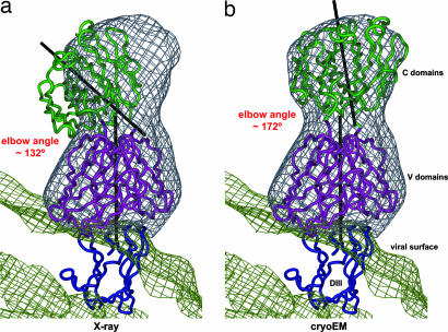
Fig. 4.
Fit of Fab E16 into the cryo-EM difference density (gray). (a) Crystal structure of the Fab E16+DIII complex shown after superpositioning of the x-ray DIII portion (blue) onto DIII-C of the fitted WNV E protein. The variable domains are shown in magenta; the constant domains are in green. The black axes represent the pseudodyads between light and heavy chain for the variable and the constant parts. (b) As in a, but the constant domains were adjusted to fit to the cryo-EM density, resulting in an increase of the elbow angle by 40°.

