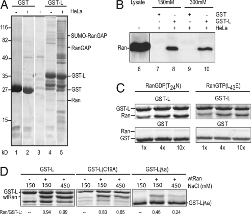
Fig. 3.
Leader–Ran interactions. (A) Recombinant GST or GST-L, bound to glutathione beads, was reacted with HeLa extracts (lanes 2 and 5) or with buffer (lanes 1 and 4). Bound protein was fractionated by SDS/PAGE and then visualized after staining. Control beads without recombinant proteins were treated in parallel (lane 3). (B) GST or GST-L beads were incubated with HeLa extracts as in A. Before boiling, the beads were washed with 150 mM NaCl (lanes 7 and 8) or 300 mM NaCl (lanes 9 and 10). After SDS/PAGE, the bands were visualized by Western assay by using anti-Ran polyclonal antibodies. Unfractionated cell extract (lane 6) was included as a Ran marker. (C) GST or GST-L beads were incubated with the indicated molar equivalents of recombinant Ran(T24N) or Ran(L43E). Extracted protein was fractionated by SDS/PAGE and then visualized by staining. (D) GST-L, GST-L(C19A), or GST-L(Δa) beads were incubated with (wild-type) Ran (1:4) as in C. The beads were washed with 150 mM or 450 mM NaCl as in B before the bound proteins were extracted, fractionated by SDS/PAGE, and visualized by staining. Relative band intensities were captured (ImageQuant) and are presented as the ratio of Ran/GST-L (or its derivatives) on the gels.

