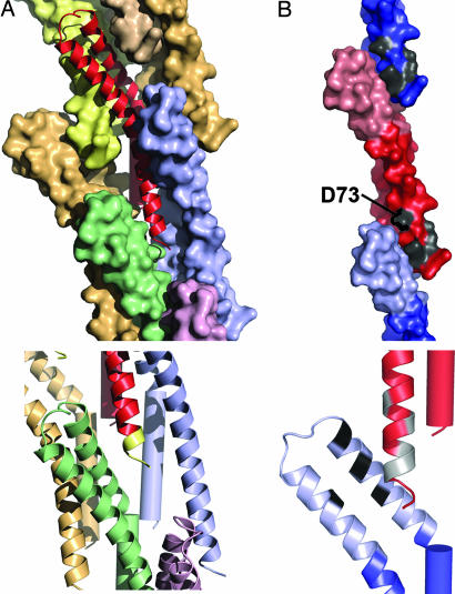
Fig. 3.
Interactions between subunits in the assembled needle. (A) A monomer of MxiH (ribbon diagram, red) is surrounded by seven identical subunits (shown as colored surface representations) within the needle assembly (Upper). The C terminus of MxiH, magnified (Lower), with the five C-terminal residues colored yellow, makes direct contact with three surrounding monomers (shown as ribbon diagrams in blue, green, and purple). (B) The tail region of MxiH (blue for the monomer above and red for the central monomer) contacts the head region of the monomer below (light red for the central monomer and light blue for the monomer below). Mutations that cause severe defects in hemolysis/invasion despite normal needle assembly (11) are shown in gray (D73A, D75A, I78A, I79A, and Q80A) (Upper). The magnification of the interface (as a ribbon diagram) (Lower) is rotated to show the patch of residues (black, L30, L34, A38, and Y50) on the head (light blue) that contact the residues listed above (colored gray on the red ribbon diagram).

