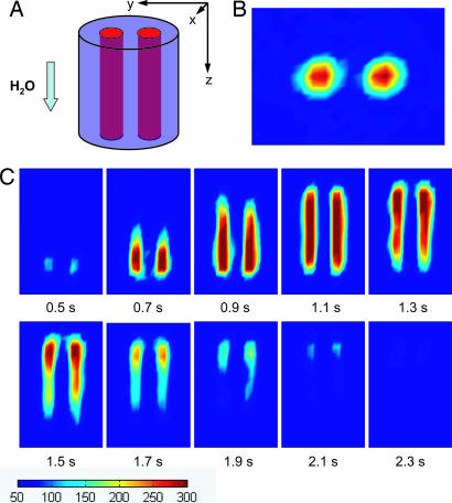
Fig. 3.
Magnetic resonance images. (A) The encoding volume. The two channels are 3.2 mm in diameter and 25 mm long each, with a center-to-center spacing of 5.1 mm. (B) Image of the cross-section of the encoding volume perpendicular to the flow (xy plane) at t = 1.1 s. (C) Time-resolved images in the yz plane. Measurements were obtained with a time interval of 0.1 s. All of the images are color-mapped at the same scale, as indicated below the images. The total experimental time for these flow images is 12 h, which is dominated by the waiting time between measurements to allow the sample from the previous measurement cycle to clear the system. The overall time will be reduced to minutes with shorter travel distances.

