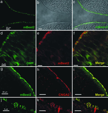
Fig. 3.
Localization of mBest2 on the sensory cilia of OSNs. (a and c) OE (bright field in b) labeled with affinity-purified anti-mBest2 polyclonal antibody (green) is shown as the fluorescence signal (a) or a digital addition of the fluorescence and bright-field images (c). mBest2 is located at the luminal surface of the sensory epithelium. (d–f) Double staining for mBest2 (red) and OMP (green). mBest2 was located at the end of dendrites of mature OSNs. (g–i) Double staining for mBest2 (green) and CNGA2 (red). Label is shown as the red fluorescence channel digitally combined with the green channel. mBest2 and CNGA2 colocalized at the luminal surface of OE. (j–l) Higher magnification of the OE as in g–i. mBest2 was expressed on cilia of OSNs and colocalized with CNGA2.

