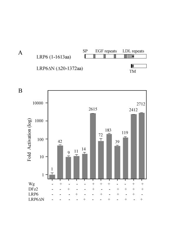
Figure 4.
LRP6 and LRP6ΔN stimulate the signaling pathway. (A) Diagram of the full-length LRP6 and its truncated construct LRP6ΔN with deleted aminoterminus. SP represents the signal peptide and TM denotes the transmembrane domain. (B) S2 cells were transfected with the indicated expression plasmids. Fold activation values were measured relative to the levels of luciferase activity in cells transfected with empty vectors and normalized by Renilla luciferase activities. These values are plotted as a log function. All experiments were done in triplicate; error bars represent the standard deviations.

