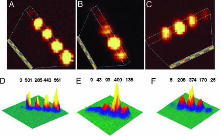
Fig. 2.
Confocal Raman microscopy images of gapped nanowire structures functionalized with MB. (A–C) Two-dimensional Raman images corresponding to the structures shown in Fig. 1 D (A), E (B), and F (C). (D–F) Three-dimensional Raman images. A Inset, B Inset, and C Inset are schematic representations of the structures being imaged. Peak intensities are in arbitrary units.

