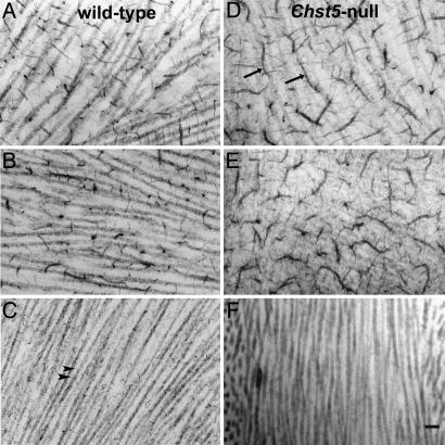
Fig. 5.
Electron micrographs of the corneal stroma in WT (Left) and homozygous Chst5-null mice (Right), showing PGs stained with Cupromeronic blue after incubation in buffer (A and D), keratanase (B and E), and chondroitinase ABC (C and F). Small collagen-associated PG filaments remaining after chondroitinase ABC digestion in the WT cornea (C, arrowheads) represent sulfated KSPGs. These are not present in Chst5-null cornea (F). Abnormally large, caterpillar-like PGs (arrows), not present in the WT stroma (A), are evident in the Chst5-null cornea (D). These are susceptible to chondroitinase ABC (F), but not to keratanase (E), digestion pointing to a significant CS/DS component. (Scale bar: 300 nm.)

