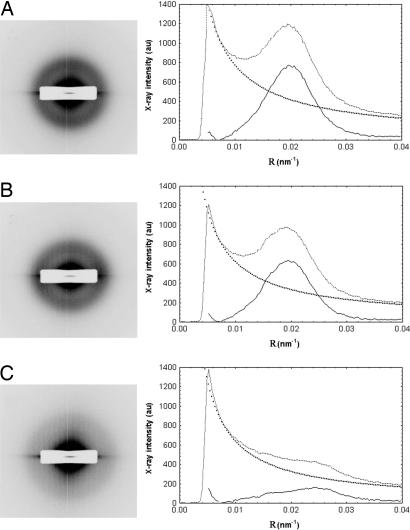
Fig. 6.
Synchrotron x-ray fiber diffraction patterns from the corneas of WT (A), heterozygous (B), and homozygous (C) Chst5-null mice revealing a more diffuse collagen interfibrillar x-ray reflection in the homozygous situation. Background-subtracted peaks in the x-ray intensity profiles from the patterns confirm the diffuseness of the interfibrillar reflection and enable calculation of the mean center-to-center collagen interfibrillar Bragg spacing.

