Fig. 3.
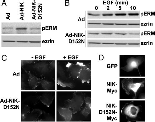
NIK activity is necessary for phosphorylation of ERM proteins by EGF. (A) Immunoblots for pERM proteins (Upper) and ezrin (Lower) in lysates of quiescent MTLn3 cells infected with Ad, Ad-NIK, or Ad-NIK-D152N are shown. (B) Time-dependent phosphorylation of ERM proteins with EGF is indicated by immunoblotting for pERM proteins and ezrin in lysates of MTLn3 cells infected with Ad or Ad-NIK-D152N. (C) Immunolabeling with anti-pERM antibodies of MTLn3 cells infected with Ad or Ad-NIK-D152N in the absence (−EGF) or presence of 25 nM EGF for 5 min (+EGF) indicates pERM proteins are predominantly in lamellipodia in Ad-infected cells with EGF. (D) GFP fluorescence (Top) and anti-Myc immunolabeling of cells expressing NIK-Myc (Middle) or NIK-D152N-Myc (Bottom) are shown. (Scale bar: 10 μm.)

