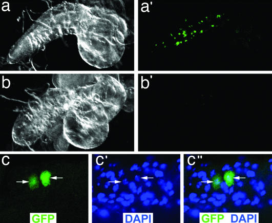
Fig. 3.
PAX7-FKHR cells enter the larval CNS. (a and a′) A dark field (a) and GFP fluorescent (a′) image of a PAX7-FKHR (MHC-Gal4, UAS-GFP, UAS-PAX7-FKHR) CNS organ with GFP-positive tissue. (b and b′) A dark field (b) and GFP fluorescent (b′) image of a wild-type (MHC-Gal4, UAS-GFP) CNS organ. No GFP-positive cells are identified, although dim, nonspecific, small foci of background autofluorescence can be seen. (c–c″) Confocal images from a GFP-positive PAX7-FKHR larval CNS organ. The white arrows highlight the individual nuclei that correspond to the GFP-positive cells. (Magnifications: c–c”, ×126.)

