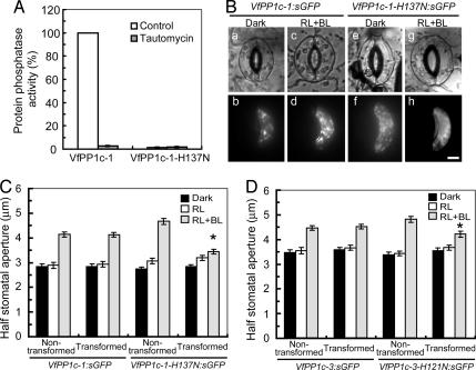
Fig. 2.
Inhibition of blue light-dependent stomatal opening by expression of mutant forms of PP1c in guard cells. (A) Protein phosphatase activity of VfPP1c-1 and VfPP1c-1-H137N prepared from E. coli cells. Phosphatase activity was measured by using [32P]-labeled myelin basic protein in the absence (control) or presence of 10 nM tautomycin. Each value was expressed as percentage of VfPP1c-1 (55,498 ± 2,914 nmol·min−1·mg−1 protein). Data are means ± SE (n = 3). (B) Images of guard cells in the bright field (a, c, e, and g) and fluorescence microscope (b, d, f, and h). Typical cases of guard cells transformed with VfPP1c-1:sGFP (a–d) or VfPP1c-1-H137N:sGFP (e–h) are shown. Gene constructs were introduced into Vicia guard cells by particle bombardment. Epidermal strips were detached and preincubated for 2.5 h in darkness, and then illuminated by red light (RL, 150 μmol·m−2·s−1) with or without blue light (BL, 10 μmol·m−2·s−1) for 2.5 h. (Scale bar, 10 μm.) (C and D) Half stomatal apertures measured next to transformed and nontransformed guard cells. Results are means ± SE (n = 45, pooled from triplicate experiments) from VfPP1c-1:sGFP or VfPP1c-1-H137N:sGFP (C) and VfPP1c-3:sGFP or VfPP1c-3-H121N:sGFP (D) transformants. Asterisks indicate significant differences between nontransformed and transformed guard cells (P < 0.01).

