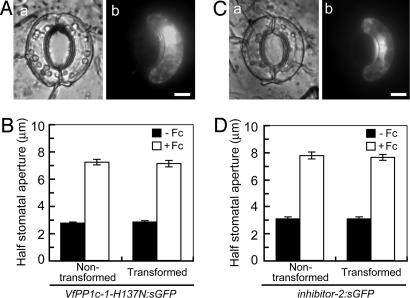
Fig. 4.
Response to fusicoccin of guard cells transformed with PP1c mutant and inhibitor-2. (A and C) Images of guard cells under bright field (a) and fluorescence conditions (b). Typical guard cells transformed with VfPP1c-1-H137N:sGFP (A) and inhibitor-2:sGFP (C) are shown. Epidermal strips were preincubated for 2.5 h in darkness before fusicoccin (10 μM) was added; images were taken after further incubation for 2.5 h in darkness. (Scale bars, 10 μm.) (B and D) Determination of half stomatal apertures of guard cells transformed with VfPP1c-1-H137N:sGFP (B) or inhibitor-2:sGFP (D). Data are means ± SE (n = 45, pooled from triplicate experiments).

