Fig. 5.
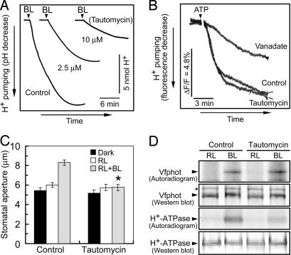
Effects of tautomycin on H+ pumping, stomatal opening, and the phosphorylation of Vfphots and the plasma membrane H+-ATPase in response to blue light. (A) Inhibition of blue light-dependent H+ pumping in Vicia guard cell protoplasts by tautomycin. Guard cell protoplasts were preincubated for 30 min under the red light (RL, 600 μmol·m−2·s−1), and then were exposed to 10 μM tautomycin for 2 h. Afterward, a pulse of blue light (BL, 100 μmol·m−2·s−1, 30 s) was applied at the times indicated by vertical arrowheads. (B) Effect of tautomycin on ATP-dependent H+ pumping in microsomal vesicles from guard cell protoplasts. The basal reaction mixture (250 μl) contained membrane vesicles (10 μg of protein), 10 mM Mops-KOH (pH 7.0), 0.25 M mannitol, 5 mM MgCl2, 1 mM EGTA, 50 mM KNO3, 5 μg·ml−1 oligomycin, and 1 μM quinacrine. Vanadate at 100 μM and tautomycin at 10 μM were added. ΔF/F, change in fluorescence divided by the initial fluorescence. (C) Inhibition of blue light-dependent stomatal opening by tautomycin. Epidermal strips were preincubated for 2.5 h in darkness with 10 μM tautomycin, and then illuminated by red light (RL, 150 μmol·m−2·s−1) with or without blue light (BL, 10 μmol·m−2·s−1) for 2.5 h. Data represent means ± SE (n = 65, pooled from triplicate experiments). Asterisk indicates significant difference between control and tautomycin treatments (P < 0.01). (D) Levels of phosphorylation and the amounts of Vfphot and the H+-ATPase. Guard cell protoplasts were treated as described for Fig. 5A with [32P]orthophosphate. Autoradiography was carried out on Vfphots and H+-ATPase, which were isolated by immunoprecipitation from 200 and 100 μg of guard cell proteins, respectively. Western blotting of Vfphot and the H+-ATPase was performed by using polyclonal antibodies raised against to individual protein. Asterisk indicates a nonspecific protein. Experiments repeated on two occasions gave similar results.

