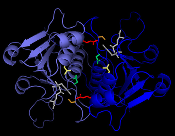
Figure 11.
Three-dimensional model of the YfhQ dimer in complex with AdoMet. Both monomers are shown in the cartoon representation, in different shades of blue. AdoMet molecules (white) and residues predicted to be involved in catalysis in YfhQ and conserved in other ribose MTases (see Figure 7) are shown in the wireframe representation in different colors (N17 in yellow, R23 in red, S142 in orange, and N144 in green). The coordinates of the model are available from the corresponding author upon request.

