
Figure 1.
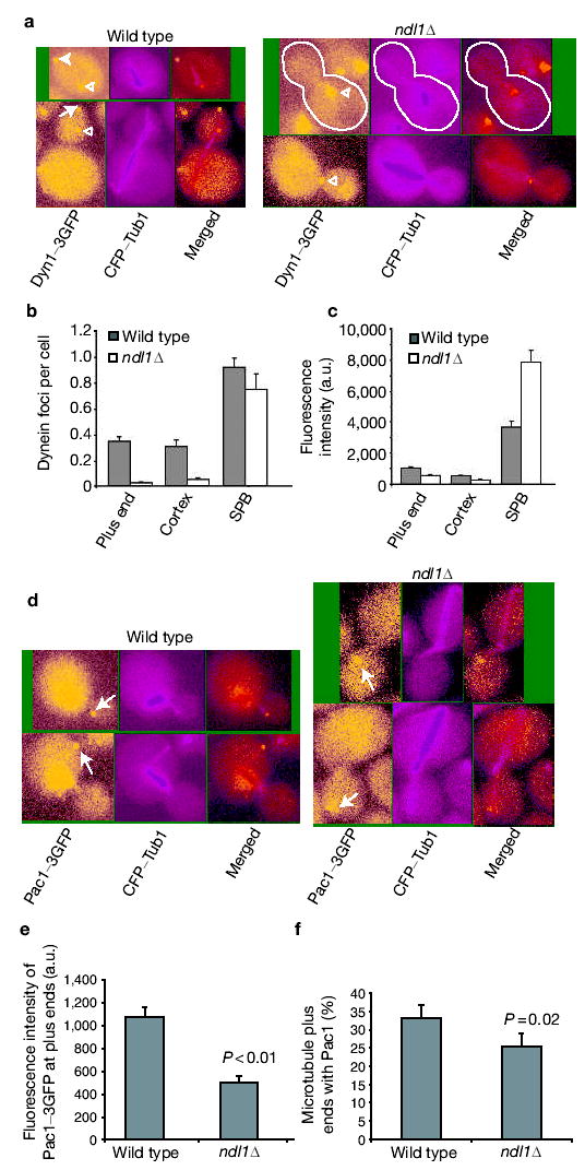
Ndl1 is required for efficient Dyn1 and Pac1 targeting to the plus ends. (a) Colocalization of Dyn1–3GFP and CFP–Tub1. Dyn1–3GFP appears at the microtubule plus end (arrow), the cortex (solid arrowhead) and the SPB (open arrowhead) in wild-type cells. In an ndl1Δ mutant, most Dyn1– 3GFP is present at the SPB. Cell shape is outlined in white. (b) Frequency of Dyn1–3GFP foci at plus ends, cortex and SPB in wild-type and ndl1Δ cells based on dual-colour images of Dyn1–3GFP and CFP–Tub1. Error bars represent standard error (n > 300). (c) Intensity of Dyn1–3GFP fluorescence intensity at the plus end, cortex and SPB in wild-type and ndl1Δ cells. Error bars represent standard error (n = 15). (d) Pac1–3GFP is present at the plus end of microtubules (arrow) and also in the nucleus. In an ndl1Δ mutant, Pac1–3GFP appears at microtubule plus ends without accumulation in the nucleus. (e) Intensity of Pac1–3GFP fluorescence at plus ends of microtubules. Error bars represent standard error (n = 15). (f) Percentage of cytoplasmic microtubules with Pac1–3GFP at the plus end (based on dualcolour images of Pac1–3GFP and CFP–Tub1) in wild-type and ndl1Δ cells. Error bars represent standard error (n > 50).