
Figure 4.
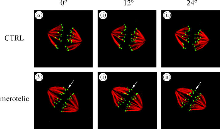
Projection views at three different angles through 3D reconstructions of two metaphase PtK1 spindles (a and b) that were fixed and immunofluorescently labelled with CREST antibodies to the kinetochore (green) and tubulin antibodies to stain kinetochore fibre microtubules (red). Cells had progressed normally through mitosis to metaphase before fixation. Note the cell in b has a merotelic kinetochore shifted toward the equator in between the other pairs of sister kinetochores (arrows). From Cimini et al. (2003).