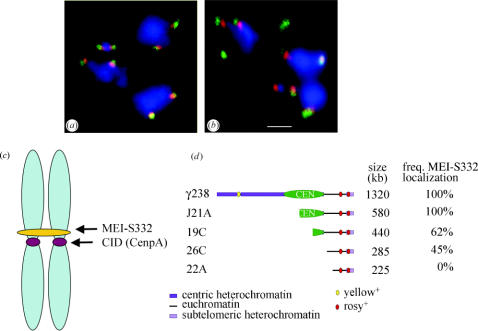
Figure 1.
MEI-S332 localizes adjacent to the kinetochore and binding is dependent on a functional centromere. (a) MEI-S332 (red) localizes adjacent to the outer kinetochore protein ZW10 (green) at the centromeres of bivalents in prometaphase I Drosophila spermatocytes. DNA is stained in blue. (b) MEI-S332 (red) also localizes adjacent to rather than coincident with dynein (green). MEI-S332 is closer to the DNA (blue) than this outer kinetochore protein. Photomicrographs in a and b are courtesy of Jacqueline Lopez; scale bar is 1 μm. (c) In spread chromosomes from mitotic Drosophila S2 culture cells, MEI-S332 is observed localized in a band on one side of the centromere, marked by localization of CID (Drosophila CenpA; Blower & Karpen 2001). (d) Deletion derivatives of a minichromosome were used to map the chromosomal sites needed for MEI-S332 localization. MEI-S332 localization was analysed by immunofluorescence in prometaphase I spermatocytes. The frequency of localization refers to the percentage of prometaphase I spermatocytes in which MEI-S332 could be detected on the minichromosome. The functional centromere region is designated by the green oval. Although the derivatives 19C, 26C and 22A are missing the centromere and are transmitted poorly, they nevertheless organize a kinetochore. The outer kinetochore protein ZW10 is always detectable on these minichromosomes. In contrast, the frequency with which MEI-S332 could be observed diminished in derivatives lacking a fully functional centromere, and MEI-S332 was not detected on the 22A derivative. Panel d is adapted from Lopez et al. (2000).

