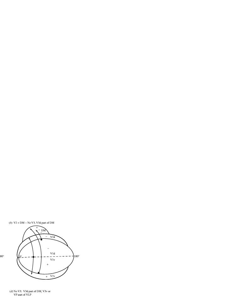
Figure 1.
Different proposals for how visual cortex in the region of V3 is organized. (a) Traditional proposals stemming from the descriptions of Cragg (1969) and Zeki (1969) include a V3 with a dorsal half, V3d, representing the lower visual quadrant, and a ventral half, V3v, representing the upper visual quadrant. The second visual area, V2, is split along the zero horizontal meridian (HM) to border primary visual cortex, V1, along the representation of the zero vertical meridian (VM). Area V3 borders much of V2, roughly as a mirror image of V2. V1 projects to retinotopically matched sets in both V2 and V3, as well as other areas including the dorsomedial visual area, DM, possibly equivalent to V3A. Sites along the outer border of V1 correspond to the VM and project to the outer border of V3, while sites along the midline of V1 correspond to the HM and project to the V2/V3 border. In the original portrayals of V3 of Cragg (1969) and Zeki (1969), V2 and V3 were continuous and of equal size. It now appears likely that in at least some primates, V3 has separated dorsal (V3d) and ventral (V3v) halves, and V3 is smaller than V2 (e.g. Kaas & Lyon 2001). (b) An alternative proposal is that a number of visual areas border V2, and there is no V3 (Allman & Kaas 1974). In this view, much of V3d is subsumed in DM, and projections of V1 that were considered to be to V3d are instead to DM (Lin et al., 1982). Because the evidence for V1 projections to V3v was uncertain, the region of V3v was considered part of another visual area (a combination of ventroposterior, VP, and ventroanterior, VA, visual areas (see Newsome & Allman 1980). (c) Perhaps the dominant theory today is that V3d is a visual area by itself even though it represents only the lower visual quadrant. The V3v region is also considered a separate visual area, the ventroposterior area, VP, which represents only the upper visual quadrant. This theory (as well as those in (b) and (d)) was supported by the erroneous conclusion that V3v does not have connections with V1 (Weller & Kaas 1983; Van Essen et al. 1986). (d) Another possibility is that much of V3d is part of DM (as in (b) above), while V3v is part of a larger area, the ventrolateral posterior area (VLP). The dorsolateral complex (DL or V4) of monkeys has been divided into rostral (DLr) and caudal (DLc) areas (see Stepniewska & Kaas 1996) and DLc has been combined with V3v/VP to form VLP by Rosa & Tweedale (2000). They have also combined the territory of DLr with the region of VA or V4v to form the ventrolateral anterior area, VLA.

