
Figure 3.
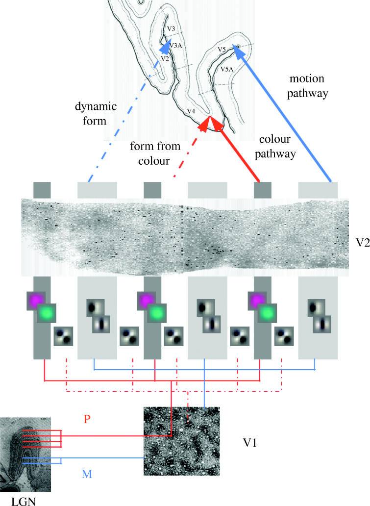
Schematic adapted from Zeki (1993) summarizing the functional segregation of processing pathways and the relationship of simulated RFs to the stripe structures in V2. LGN, lateral geniculate nucleus; P, parvocellular pathway; M, magnocellular pathway. These RFs were obtained by minimizing the free energy of a model neuronal system when exposed to moving natural scenes. See Friston (2000) for details.