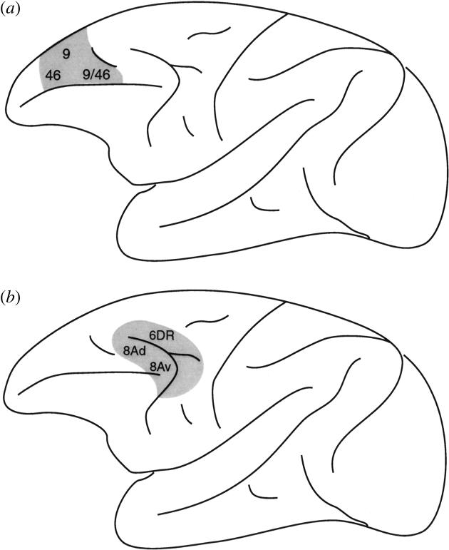
Figure 5.

Schematic illustration of (a) the mid-dorsolateral (MDL) prefrontal lesion and (b) the caudal dorsolateral prefrontal lesion, which involved the cortex within and around the dorsal arcuate sulcus, i.e. the periarcuate (PA) region. These lesions in the monkey were used to study fundamental differences in function along the rostral–caudal axis of lateral frontal cortex. The numbers refer to the architectonic areas involved in these lesions.
