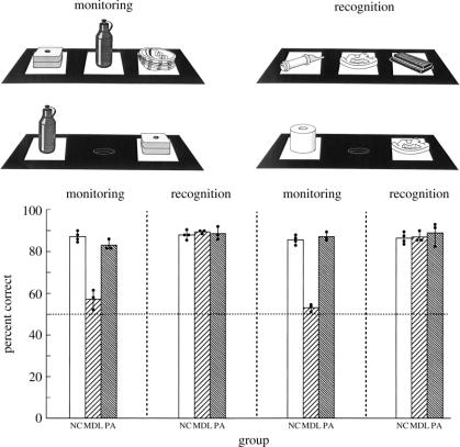
Figure 7.
(Upper panel): schematic diagram of the experimental arrangement in the self-ordered monitoring working memory condition and the recognition memory condition administered to the monkeys. The upper displays illustrate the presentation trials and the lower displays the test trials in both the monitoring and recognition conditions. (Lower panel): postoperative performance of animals with mid-dorsolateral frontal lesions (MDL), animals with periarcuate lesions (PA) and normal control animals (NC). The mean per cent correct performance over the four postoperative testing blocks (20 days of testing per block) is shown. Solid circles indicate the scores of individual animals in each group. In the monitoring condition, the animals with MDL lesions were severely impaired, whereas the animals with PA lesions performed as well as the NC animals. Both groups with lesions performed as well as the normal control animals in the recognition memory condition. Data from Petrides (1991).

