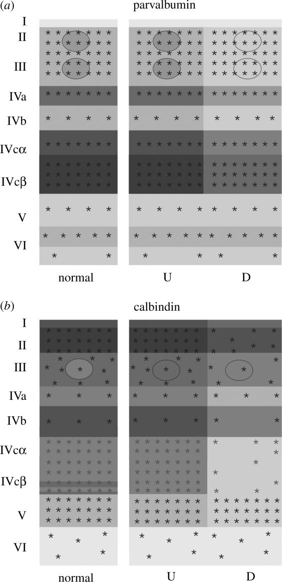
Figure 5.

Schematic of the laminar distribution of cell bodies (asterisks) and neuropils (greys) in coronal sections of V1, stained for parvalbumin (a) and calbindin (b), after massive retinal laser lesions. Schematics of the stains are illustrated for normal and for deprived (D) and undeprived (U) columns.
