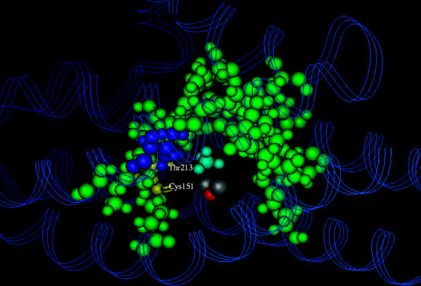
Figure 5.
A computational model of the possible binding site for methane in the hydroxylase of sMMO. The hydrophobic residues forming the ‘horseshoe-shaped’ pocket are shown in green. The binuclear iron centre is in silver/grey, and the bound methane molecule is shown in blue (George et al. 1996).

