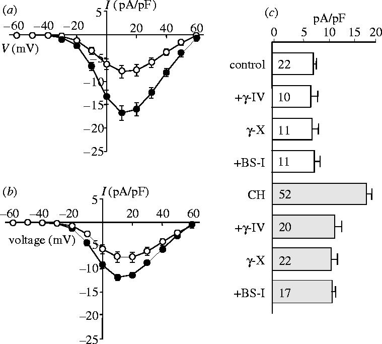
Figure 5.

Hypoxia enhances Ca2+ channel currents in HEK-293 cells stably expressing the human cardiac L-type Ca2+ channel α1C subunit. (a) Mean (+s.e.m. bars) current density versus voltage relationships obtained from control cells (open symbols, n=12) and from cells cultured in a hypoxic atmosphere of 2.5% O2 for 24 h prior to recording (solid symbols, n=15). (b) Mean current density (+s.e.m. bars) versus voltage relationships obtained from control cells (open symbols, n=9) and from cells pretreated for 24 h with 20 nM AβP1–40 (solid symbols circles, n=18). (c) Bar graph showing mean (+s.e.m.) current density (determined at a test potential of +20 mV) in control cells (open bars) and in hypoxically cultured (CH) cells (shaded bars) exposed to various secretase inhibitors (the β-secretase inhibitor, BS-I (30 nM), the γ-secretase inhibitor, γ-IV (2.5 μM) and the γ-secretase inhibitor, γ-X (100 nM), as indicated). Values in bars indicate n numbers.
