
Figure 2.
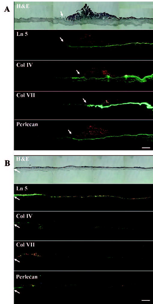
Overview of BM formation of limbal corneal explants cultured on iAM or dAM for 4 weeks. When an explant was cultured on iAM for 4 weeks (A), Ln-5, Col IV, Col VII, and perlecan were strongly expressed as a continuous band underneath the epithelial outgrowth, but decreased and became negative farther out from the outgrowth’s leading edge (arrow). In contrast, when an explant was cultured on dAM for 4 weeks (B), Ln-5, Col IV, Col VII, and perlecan started to deposit in a linear-segmented band close to the leading edge of the outgrowth (arrow) and showed a spotted and cytoplasmic staining in the major part of the outgrowth. Bar, 200 μm.