
Figure 7.
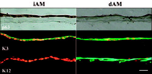
Differentiation markers expressed by outgrowth epithelial cells expanded on iAM or dAM for 4 weeks. There was more nuclear staining of p63 on iAM than dAM. K3 was negative or weakly positive in basal epithelial cells on iAM, but was strongly positive in all outgrowth epithelial cells on dAM. K12 was weakly positive on iAM, whereas almost all cells were strongly positive on dAM. Bar, 50 μm.