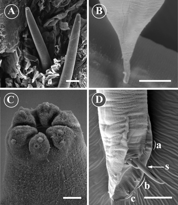
Figure 5.
Morphology of Pelodera strongyloides from SEM. A) Two Pelodera strongyloides larvae within a hair follicle with clearly discernible lateral alae and a striated cuticle can be observed intermingling with keratin. Scale bar = 20 μm. B) The posterior end of a female Pelodera strongyloides. The tail possesses a clearspine-like extension. Scale bar = 10 μm. C) The anterior end of an adult Pelodera strongyloides. Oral opening is surrounded by six well-defined lips. Distinct papillae are present on the lips. Scale bar = 2 μm. D) The posterior end of a male Pelodera strongyloides. The scanning electron micrograph shows a copulatory bursa with its papillae: precloacal papillae (a) the anterior group of postcloacal papillae (b) and the posterior group (c) of three postcloacal papillae. Spicules (s) are protruding from the cloaca. Scale bar = 20 μm.

