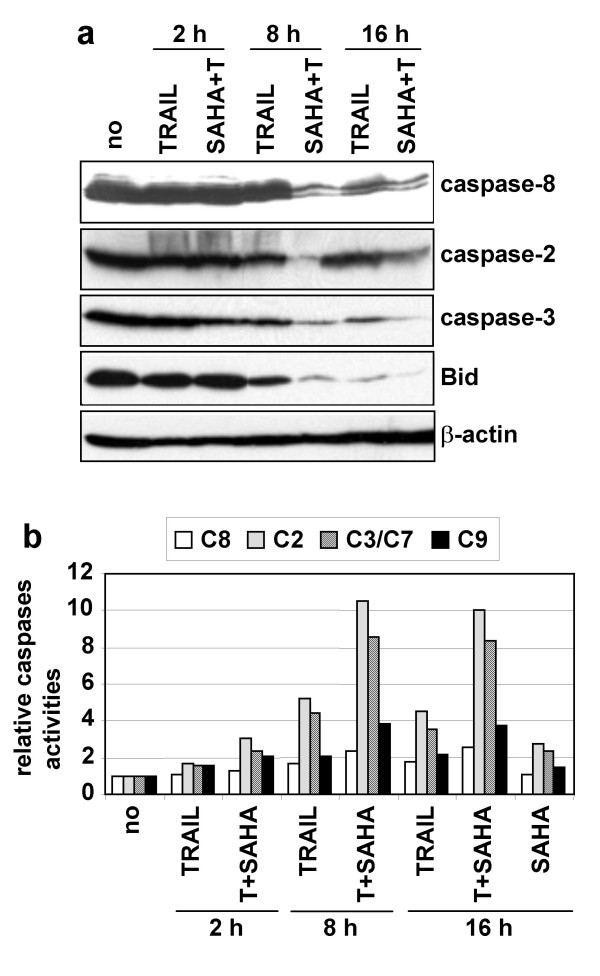
Figure 4.

Time-course analysis of caspases activation. A SH-EP cells were unstimulated (no) or stimulated with TRAIL (25 ng/ml) and M2 (125 ng/ml) (T) alone or in combination with SAHA (2.5 μM) for 2, 8 or 16 h. Whole cell extracts were analysed by immunoblotting for the cleavage of caspases-8, -2, -3, and Bid. β-actin was used as loading control. B Time-course analysis of caspases activation. Caspases-8, -2, -3/7 and -9 activities were measured in lysates of SH-EP cells treated as in a) by measuring the hydrolysis of their respective caspases colorimetric substrates. Caspases activities of stimulated cells, relative to unstimulated cells are indicated.
