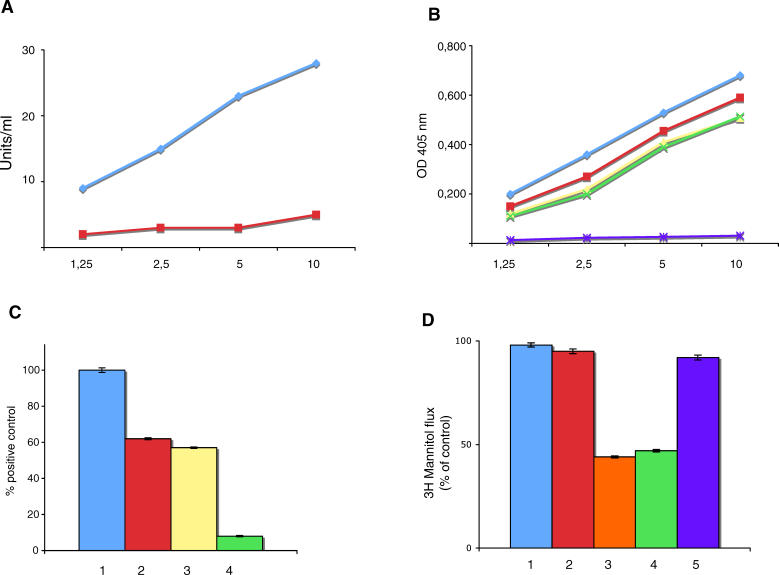
Figure 10. Features of Rabbit Anti-VP-7 Antibodies.
(A) Binding of rabbit anti-VP-7 antibodies to tTG (blue line). Red line indicates pre-immune rabbit serum.
(B) Binding of rabbit anti-VP-7 antibodies to VP-7 peptide (blue line), celiac peptide (red line), desmoglein peptide (yellow line), and TLR4-peptide (green line). Purple line indicates binding to the irrelevant control peptide.
(C) TLR4 activation by LPS (100 ng/ml) (1), affinity-purified human anti-celiac peptide antibodies (2), rabbit anti-VP-7 antibodies (3), and pre-immune rabbit serum (4). Results are expressed as percentage of positive control, where the positive control is the OD value obtained upon stimulation of TLR4 transfected cells with 100 ng/ml LPS (maximal concentration used).
(D) Confluent T84 monolayers were treated for 3 h with control normal human Ig (1), affinity-purified human antibodies to the irrelevant control peptide (2), affinity-purified human anti-celiac peptide antibodies (3), rabbit anti-VP-7 antibodies (4), and pre-immune rabbit serum (5).

