
Figure 2.
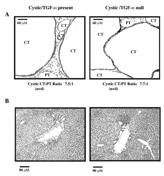
Histologic appearance of d-21 kidneys and livers from cystic bpk mice with TGF-α present or absent. (A) Serial kidney sections from cystic mice with TGF-α (Cystic/TGF-α present) or without TGF-α (Cystic/TGF-α null) were stained with biotinylated Dolichos biflorus agglutinin (DBA), a CT marker or biotinylated Lotus tetragonolobus (LTA), a PT marker and counter-stained with hematoxylin as described in the text. Representative kidney histologies are shown. CT cysts (DBA+)/LTA− are indicated by dark staining and predominate in both groups studied. A minority of cystic PT (DBA−/LTA+) is present. The cystic CT:PT ratio is indicated below each panel. Neither the overall severity of cystic CT disease nor the appearance or proportion of cystic PT appears to be modulated by the presence or absence of TGF-α. CT, collecting tubule cyst; PT, proximal tubule cyst. Original magnification 20×. (B) Serial liver sections from cystic mice with TGF-α (Cystic/TGF-α present) or without TGF-α (Cystic/TGF-α null) were stained with hematoxylin as described in the text. Representative liver histologies are shown and demonstrate that the severity of bile duct ectasia and proliferation was qualitatively similar in mice with versus without TGF-α. Original magnification 20×.