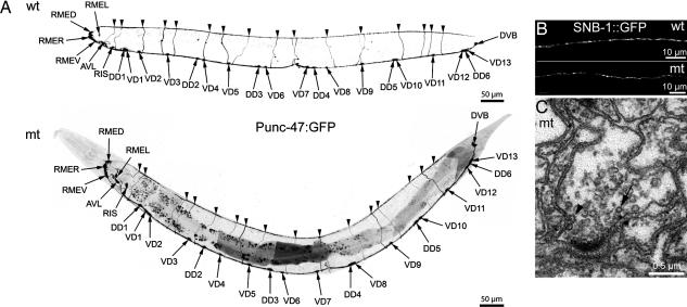
Fig. 4.
Neuronal architecture is normal in the TVP triple mutant. (A) Fluorescence micrographs (negative image) of the distribution of GABA neurons expressing GFP under the control of the unc-47 promoter in wild-type (wt) and mutant (mt) backgrounds (strains EG1285 and BJ1, respectively). (B) Fluorescence micrographs of SNB::GFP in the dorsal nerve cord in wild-type and mutant backgrounds (strains BJ22 and BJ28, respectively). (C) Electron microscopy of a synapse in the ventral nerve cord of a triple TVP mutant worm (strain EG2960). Note the presence of clustered synaptic vesicles close to a typical presynaptic specialization, the occurence of clathrin-coated vesicles (arrow), and the detection of an endocytotic figure (arrowhead).

