
Figure 3.
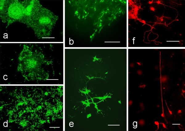
Neurons show enhanced migration patterns and neurite outgrowth when differentiating from neurospheres derived from PTPσ (-/-) mice. All images taken after 7 days of differentiation. Epi-fluorescence with the β-III tubulin antibody. A. Morphological appearance: a) Appearance of neurospheres from PTPσ(+/+) mice grown on laminin coated coverslips. Scale bar = 150 μm. b) Higher magnification of (a) to show neuronal progeny derived from the PTPσ(+/+) genotype. Scale bar = 100 μm c) Appearance of the neuronal migration from PTPσ(+/-) derived neurospheres. Scale Bar = 100 μm d) Neurons that differentiate from neurospheres from a PTPσ(-/-) mouse appear extensively distanced from the neurosphere of origin. Scale bar = 100μm. e) Neuronal differentiation from a PTPσ(+/-) neurosphere. Scale bar = 50μm. f) Long random processes characterize PTPσ(-/-) neurons. β-III tubulin immunocytochemistry. Scale bar = 50μm. g) High power view of a long process from a neuron derived from a PTPσ(-/-) neurosphere. β-III tubulin immunocytochemistry. Scale bar = 25μm.