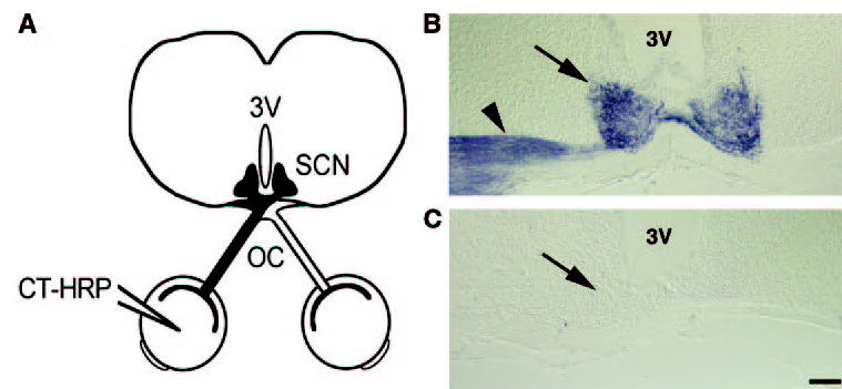
Figure 2.

Math5-null mice lack retinohypothalamic tracts. (A) CT-HRP was injected into one eye, labeling its optic nerve and projections in the brain, including the optic chiasm (OC) and suprachiasmatic nuclei (SCN). Coronal brain sections were stained with TMB to visualize RGC projections. (B) In wild-type mice, the ipsilateral RHT (arrowhead) and both SCN (arrow) were labeled, whereas the contralateral RHT was unstained. (C) In Math5−/− mice, the SCN (arrow) and the RHT were unstained. The ventral surface is at the bottom of each panel. 3V, third ventricle. Scale bar, 100 μm.
