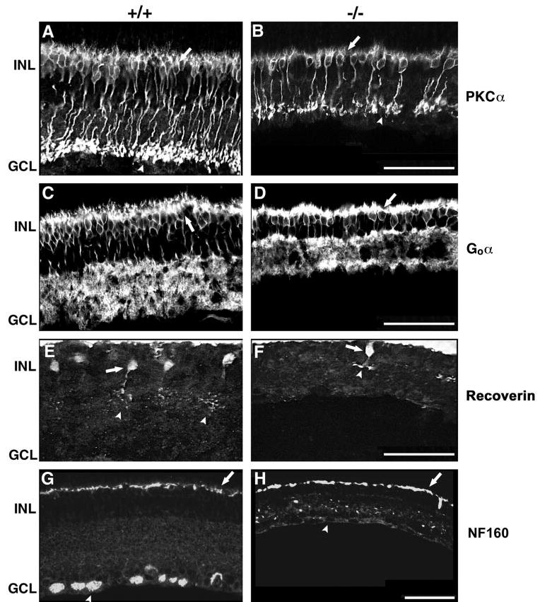
Figure 7.

Confocal immunofluorescence micrographs showing that all bipolar subtypes are decreased in Math5 mutants. (A, B) PKCα staining of rod bipolar cells. Somata (arrows) and axon termini (arrowheads) are indicated. (C, D) Goα staining of cone ON and rod bipolar cells. Arrows: Goα-positive somata. (E, F) Recoverin staining of cone OFF bipolar cells (arrows). Their axon termini are located in the OFF sublamina of the IPL (arrowheads). (G, H) Neurofilament 160-kDa staining of RGCs (arrowhead) and horizontal cells (arrows). The labeling of horizontal cells is equivalent in both retinas, but RGCs are absent in the Math5−/− animals. Scale bar, 50 μm.
