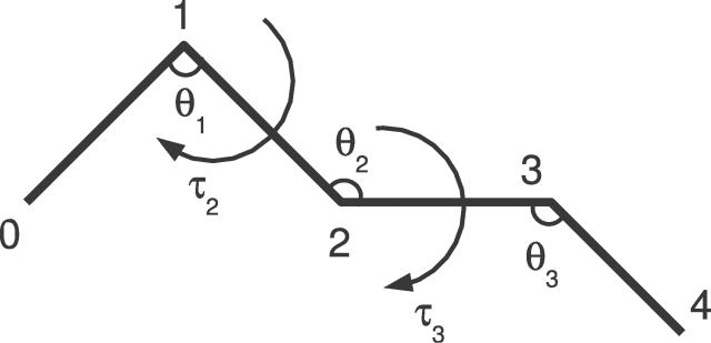
Figure 1. Schematic Representation of a Protein's Cα Backbone.
The Cα positions are numbered, and the pseudo bond angles θ and pseudo dihedral angles τ are indicated. The segment has length 5, and is thus fully described by two pseudo dihedral and three pseudo bond angles. The numbering scheme of the angles is chosen so that the angle pair (θi,τi), associated with position i, specifies the position of the Cα atom at position i + 1.

