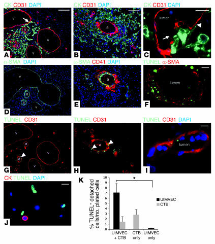
Figure 1. Human cytotrophoblasts induce arterial apoptosis during vascular remodeling in vivo.
Implants (first trimester human placental villi in Scid mouse mammary fat pads) were excised by dissection after 3 weeks and examined histologically. (A and B) Cytokeratin 7–positive cytotrophoblasts invaded murine stromal tissue, where they interacted with arteries (a) and veins (v). CD31 stain indicates endothelial cells. Arrow denotes apparent colocalization (yellow) within the vein wall. Nuclei were stained with DAPI. CK, cytokeratin. (C) Invading cytotrophoblasts displaced murine endothelial cells (arrow) and extended their cellular processes along the basal lamina (arrowhead). (D) α-SMA distribution in adjacent sections identified arteries, which had thicker tunica media than did veins. (E) Cytotrophoblast invasion disrupted arterial integrity and resulted in platelet deposition, visualized by staining for CD41. (G and H) TUNEL labeling revealed that cytotrophoblasts induced apoptosis of arterial smooth muscle and endothelial cells (arrowheads) but did not affect veins. (F and I) Staining TUNEL-labeled tissue sections for α-SMA (F) or CD31 (I) revealed that both cell types underwent apoptosis. (I) High-magnification image of a dying endothelial cell. (J and K) Cytotrophoblasts induced UtMVEC apoptosis in vitro. (J) After coculture with cytokeratin-positive cytotrophoblasts, approximately half the cytokeratin-negative UtMVECs that remained attached to the culture substrate were TUNEL labeled. (K) Coculture also increased the number of UtMVECs that underwent apoptosis and detached. CTB, cytotrophoblasts. Values are mean ± SEM from multiple experiments. *P < 0.01. Scale bars: 50 μm (A, D, and G); 40 μm (B, E, and H); 10 μm (C, F, I, and J).

