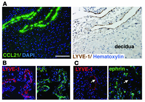
Figure 3. LYVE-1–positive vessels in the human decidua express other markers of lymphatic endothelium.
(A) Adjacent sections were analyzed. In situ hybridization with a CCL21-specific probe (green, left panel) showed that LYVE-1–positive vessels (brown, right panel) in the decidua express this chemokine. (B) Immunostaining of adjacent sections revealed the colocalization of LYVE-1 and integrin α9β1. (C) In contrast, a pan-ephrin antibody failed to label decidual lymphatic vessels (arrows), but stained invasive cytotrophoblasts (3). Scale bar: 50 μm.

