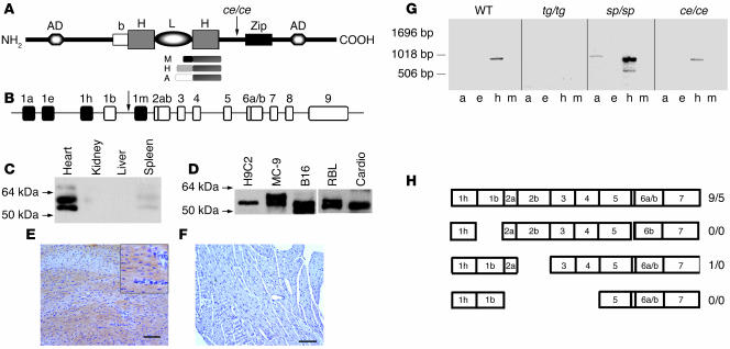
Figure 1. MITF is expressed in cardiomyocytes.
(A) Structure of MITF protein. The ce/ce mutation is a stop codon between the bHLH and the leucine zipper. b, basic domain; zip, leucine zipper. Also shown is a schematic representation of MITF protein products, with different N termini and common C terminus. (B) Genomic organization of the mi locus. tg/tg mice have an insertion of about 50 copies of a transgene incorporated into the promoter region of MITF. Filled boxes represent alternative promoters. Arrow indicates tg/tg insertion site. (C) Western blot analysis of heart, liver, kidney and spleen of normal mice. Strong expression of MITF is noted in the heart. (D) Western blot analysis of H9C2 cardiomyocytes, MC-9 mast cells, B16 melanoma cells, rat basophilic leukemia (RBL) cells, and primary culture of rat neonatal cardiomyocytes (cardio). MITF appears in cardiomyocytes at approximately the same molecular weight as in mast cells but not in melanocytes. (E and F) Immunohistochemical staining of MITF using monoclonal antibody directed against the NH2 terminus in normal (E) and tg/tg mutated (F) hearts. MITF staining is absent in tg/tg mutated hearts. Scale bars: 100 μm. Higher magnification appears in the inset. (G) cDNA from hearts of either WT or tg/tg and ce/ce MITF-mutated mice and their normal littermates (sp/sp) were amplified by PCR. Four mouse MITF alternative first exons (1a, 1e, 1h, and 1m) were used as sense primers, and exon 5 was used as antisense. Numbers on the left indicate molecular weight (bp). (H) Schematic representation of splicing patterns described for MITF. Numbers on the left of slashes represent clones including exon 6a; numbers on the right represent clones without exon 6a. Most clones are full length, including exon 6a.

